Our research focuses on the application of novel optical phenomenon for the development of advanced imaging techniques.
Research in progress
- Single molecule imaging
- Total Internal Reflection Fluorescence Microscopy (TIRFM)
- Structured Illumination Microscopy (SIM)
- Physical optics simulation
Single molecule imaging

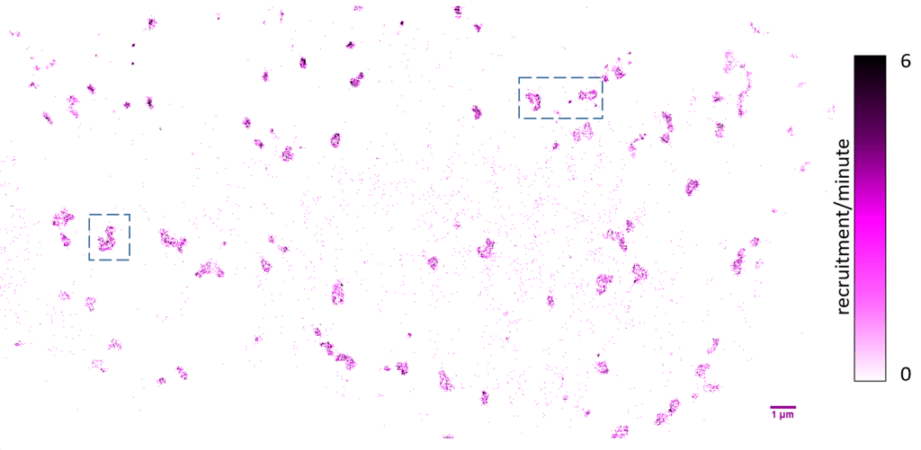
Clathrin-coated structures are the best characterized endocytic carriers, but the mechanism through which clathrin-coated plaques perform endocytic functions is still debated. We developed novel experimental and analytical approaches that allow us to dissect the mechanism of plaque growth and disassembly with high spatial and temporal resolution. We found that clathrin and its primary adaptor protein (AP2) are incorporated to the plaques at the peripheral regions. Patches of clathrin coats are internalized also from the peripheries. Our results show that the peripheral regions of plaques are active endocytic sites whereas the central regions are more stable.
Single molecule detection events and localization of AP2 on the plasma membrane. (left) acquisition of imaging AP2-GFP (middle) construction of the single molecule image by addition of localizations of each detection events (right) overlay of single molecule image and diffraction-limited image (red)

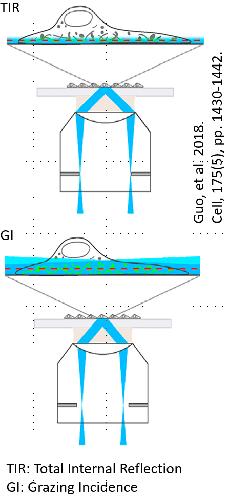
Total Internal Reflection Fluorescence Microscopy (TIRFM)
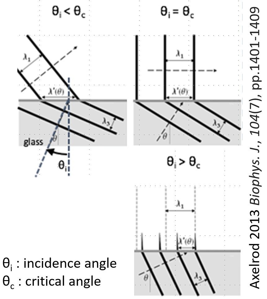
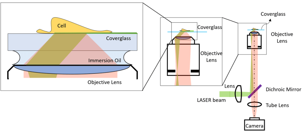
Evanescent waves are the manifestation of the boundary conditions at the interface of two distinct optical materials upon Total-Internal Reflection (TIR). They carry higher resolution spatial information. Further, they decay exponentially away from the interface, which allows confinement of illumination to the proximity of the interface and sectional illumination. The confinement of the illumination increases the imaging contrast and decreases the exposure of the sample to the laser. The incidence angle of the beam at the interface can be decreased further beyond the critical angle of TIR and hence the beam can transmit through and propagates at a small angle to the interface. At this mode a grazing incidence (GI) illumination is obtained, which can penetrate deeper into the sample and still provides a sectional illumination.
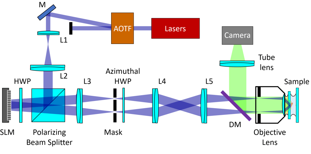
Structured Illumination Microscopy (SIM)
The amplitude of a wave propagates as a sinusoidal distribution, and when two waves Interfere the waves become a standing wave. Then the amplitude of the beams is due to the constructive and destructive interferences between the two waves. Structured Illumination Microscopy (SIM) is based on this physical principle and allows spatial confinement of the beam intensity as small as half of the wavelength. The scanning of this pattern and reconstruction of the images allows improvement of the resolution by two. SIM can be combined with other sectional illumination techniques such TIR and GI to yield improved imaging quality.

Physical optics simulation

Beam propagation can be simulated by ray or wave-based approaches or hybrid methods. Wave-based approaches can account for diffraction related effects, which are due interaction of the waves with optical features with sizes on the range of the wavelength. For simulation of the systems where diffraction related effects can be ignored, computationally more efficient ray-based simulation approaches can provide quick and accurate results. Hybrid approaches, where ray/wave-based approaches interchangeably used, offer alternative fast solutions to account for diffraction related effects.
